

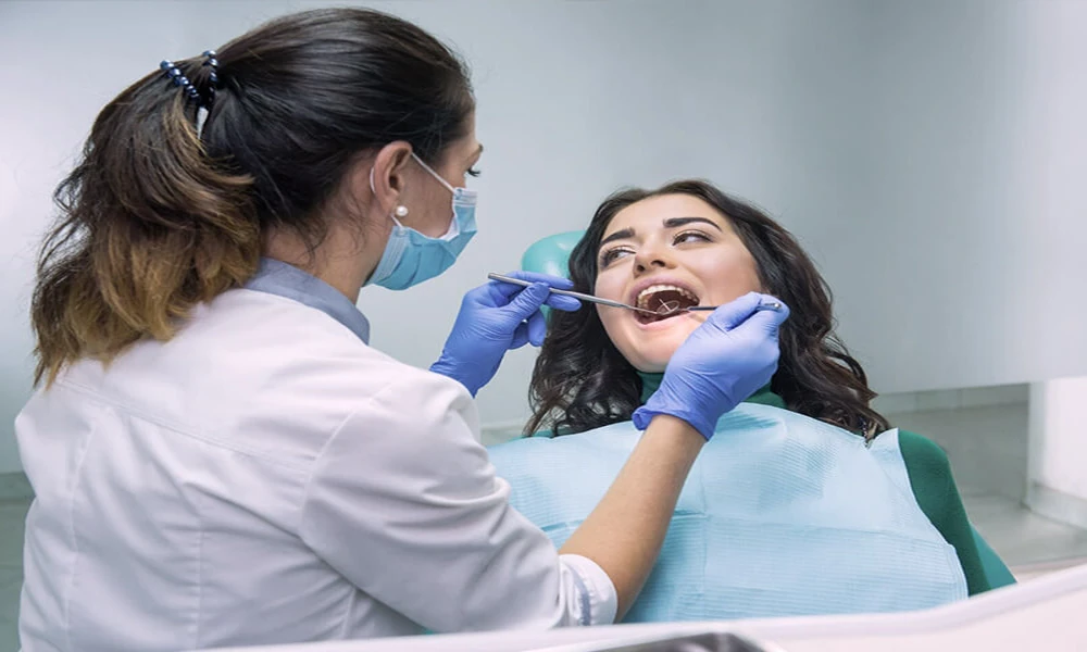
You sit in the dental chair, hear that a small spot will be “watched,” and then go home with a little knot in your stomach. That reaction makes sense. A change in the mouth can feel vague and unsettling, especially when it does not hurt and no one can promise what it means on day one. The good news is that careful tracking is part of good care. General dentists do not just glance at an area and hope for the best. They record what they see so the next visit has something solid to compare. If you also need to schedule a denture consultation in Covina, your dentist can help guide you through that process as well.
How general dentists document early lesions for future comparison comes down to consistency. They note the size, color, shape, texture, exact location, and whether the area wipes off, bleeds, feels firm, or has changed over time. Many also use photos and chart notes together. That record helps separate a harmless irritation from a spot that needs more testing or a referral.
General dentists rely on repeatable records when a lesion is not ready to diagnose on sight
Early oral lesions can be frustrating because they do not always announce themselves clearly. A small white patch near a cheek bite, a red area under a denture edge, or a flat pigmented spot on the gum may look minor at first. Some resolve when the source of irritation is removed. Some stay the same for years. Some change in subtle ways that only become clear when one visit is compared to another.
That is why documenting oral lesions for future comparison matters so much. A dentist usually starts with a written description. The note often includes the site, measured size, surface appearance, border shape, color pattern, and symptoms. If the lesion is on the right buccal mucosa opposite a certain tooth, that detail gets written down. If it is rough, ulcerated, movable, fixed, tender, or painless, that gets written down too.
Photos add another layer. A photo taken with good lighting and a consistent angle can show changes that memory misses. If a spot looks slightly larger three weeks later, the image can support that impression. If it looks the same, that matters too. This is one reason many practices include intraoral photography in a routine exam when a suspicious or uncertain area is found.
Some offices also use diagrams in the chart, marking the lesion on an oral map. That simple step helps when the area is hard to describe in words alone. If more than one provider sees you over time, the record stays clear.
According to the National Institute of Dental and Craniofacial Research oral cancer information, regular oral exams help identify abnormal areas early. The National Cancer Institute’s oral screening guidance also explains that suspicious findings may need follow up, biopsy, or referral rather than simple observation.
Changes over time often matter more than a single snapshot
A lesion that stays stable after the cause is removed can point in one direction. A lesion that enlarges, hardens, ulcerates, or shifts in color points in another. That is where tracking early mouth lesions becomes more than paperwork. It becomes a clinical timeline.
You can see why this matters in real life. Say a patient has a white patch where they often bite their cheek. The dentist smooths a sharp tooth edge, takes a photo, measures the patch, and asks the patient to return in two weeks. If the patch is gone, the record supports a likely frictional cause. If it is still there, unchanged or larger, the next step may be referral or biopsy. Without that first record, the second visit becomes guesswork.
This approach also protects patients from two common problems. The first is false reassurance. A spot that “looks probably fine” but slowly changes can be missed if no one documented the baseline. The second is unnecessary panic. Some normal variations look alarming to patients, and clear records can prevent overreaction when the area remains unchanged.
For dentists, lesion documentation in general dentist settings usually includes a follow up interval. That interval may be short if the appearance raises concern. Educational materials used in oral pathology training, such as this University of Iowa oral lesion lecture handout, reinforce the value of describing lesions in a structured way and reassessing them with intent.
Written notes, photos, and follow up each fill a different role
| Documentation method | What it captures well | Limits | Why it helps future comparison |
|---|---|---|---|
| Written chart note | Location, symptoms, texture, border, color, size | Depends on detail and wording | Creates a baseline and supports clinical decisions |
| Intraoral photo | Visual appearance, color pattern, shape | Lighting and angle can affect appearance | Shows visible change over time |
| Measurement in millimeters | Size and growth | Small errors can happen if landmarks are unclear | Turns “looks bigger” into something objective |
| Follow up visit | Progress after removing irritation or after healing time | Only works if the patient returns | Confirms whether the lesion resolves, stays stable, or worsens |
No single method does the whole job. A brief note with no photo can miss visual nuance. A photo with no size measurement can leave room for debate. A careful dentist uses more than one tool because the goal is not just to notice a lesion once. The goal is to understand its behavior.
Three steps help you take an active role in lesion follow up
1. Keep the follow up appointment. If your dentist wants to recheck an area in two weeks, a month, or three months, that timing matters. Lesions are often judged by how they change, not just how they look on one day.
2. Tell your dentist what has changed between visits. Mention pain, bleeding, roughness, numbness, growth, or whether the spot rubs against a denture, tooth, or appliance. Your day to day experience adds context that a photo cannot capture.
3. Ask what was documented. You do not need a technical lecture. A simple question helps: where is it, how big is it, what does it look like, and when should it be checked again. Clear answers reduce confusion and help you remember what to watch for at home.
Careful documentation supports earlier action when a lesion does not behave normally
Most small oral lesions are not a crisis, but they do deserve respect. Good records give your dentist a clean starting point, make follow up more reliable, and reduce the risk of missing gradual change. That is the real value behind how general dentists document early lesions for future comparison. It is steady, practical care built around evidence rather than memory.
If your dentist has asked to monitor a spot, take that plan seriously and keep the next visit on the calendar. If you notice a mouth sore, white patch, red area, lump, or pigment change that does not go away, schedule an exam with a general dentist and have it documented properly.
